LABORATORY for STRUCTURE and ULTRASTRUCTURE
MORHOMETRY and QUANTIFICATION
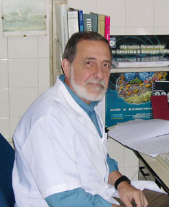
Waldemar Silva Costa, B.Sc., Ph.D.
- Biologist
- Researcher, Urogenital Research Unit
- Morphometry and Quantification
- e-mail: manogallo.c@gmail.com // cmgallo@uerj.br
Carla Braga Mano Gallo, BSc, Ph.D.
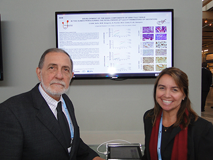
29th Annual EAU Congress in Stockholm (April - 2014)
- Best Poster -
Development of the main components of erectile tissue in the human penis during the fetal period (2nd and 3rd trimesters of gestation)
The Laboratory for Structure and Ultrastructure of the Urogenital Research Unit develops its work mainly by means of histochemical, immunohistochemical, transmission electronic microscopy and scanning electronic microscopy techniques in prostate, bladder, testicle and penis. The aim is to analyze the extracellular matrix, smooth muscle and parenchyma of these organs, performing both qualitative and quantitative analysis. Normal individuals (adults and fetuses) and the alterations that occur in congenital and acquired pathologies, benign and malignant, and during development are specially investigated. The several projects in progress employ: a) - human tissue samples from surgery, biopsy and necropsy, and b) - experimental models in animals.
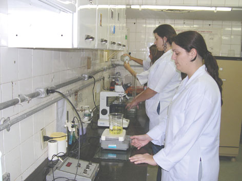
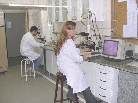
The physical facilities of the laboratory include: a) Bench space for up to six graduate and undergraduate students simultaneously; b) Basic equipment such as double boiler, heaters, bench centrifuge, centrifuge for microtubes, magnetic shakers and shakers for test tubes, diverse glassworks and various reagents, automatic pipettes, pHmeter, analytic balances, microwave oven, pipette washer, deionizer, distiller, potentiometer, etc.; c) Resources for all histochemical and immunohistochemical techniques, such as simple microscopes, microscope with polarization, digital photomicroscope; d) Olympus rotary microtome, all batteries and reagents for histology and histochemistry; e) all batteries and all antibodies correspondent to urogenital research with immunohistochemistry; f) Equipment for immunofluorescence, such as bottles of liquid nitrogen; g) Cryostat and dark room with microscope for immunofluorescence; h) Equipment for dissection of small sized animals, such as microsurgical instruments, stereoscopic microscope and optic fiber; i) Equipment for histomorphometry and stereology, Olympus microscope, Olympus camera coupled to microscope for image capture, high potency microcomputer, with specific softwares for morphometry and image analysis, including software Image Pro Plus, videomicroscopy system with color camera of image obtainment, coupled with microcomputers; j) Equipment for transmission electronic microscopy: Leika ultramicrotome with knife maker, light equipment by optic fiber, unity of block formation for electronic microscopy, and Zeiss Electronic Transmission Microscope, M-109 model; h) Scanning Electronic Microscope of low vacuum, LEO-EM 906 model, shared with other Departments of the Biomedical Center.
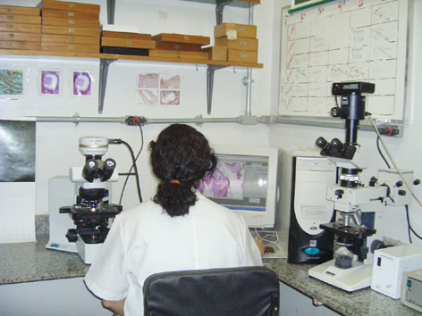
Section of immunohistochemistry
Section of computerized histomorphometry - I
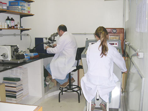
Section of computerized histomorphometry - II
Section of electronic microscopy material preparation and frozen section room
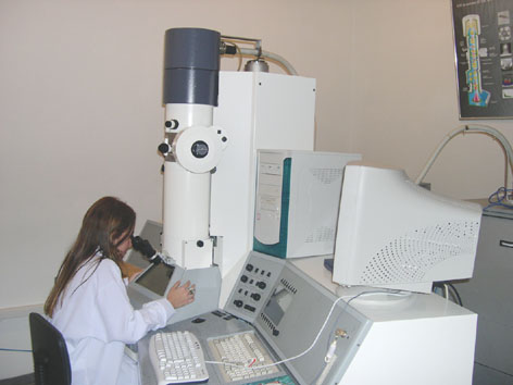
Transmission eletronic microscope, Zeiss M-190 model
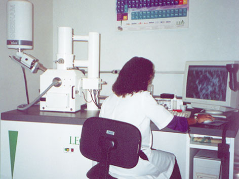
Scanning eletronic microscope of low vacuum, LEO-950 model Research Lines and Projects Currently in Progress
Extracelullar Matrix and Excretory System
- Extracellular Matrix Analysis of the Renal Calices in Normal and Dilated Collecting System.
- Extracellular Matrix and Smooth Muscle Analysis of Normal and Dilated Ureters.
Bladder - Histochemical, Immunohistochemical and Ultrastructural Study of the Urinary Bladder Wall
- Vesical Wall Structure and Ultrastructure of Rats after Hyperdistention Mediated by Liquid (In collaboration with the Federal University of São Paulo).
- Quantitative Study of the Bladder Wall Extracellular Matrix Components in Non-Obstructed and in Patients with Infravesical Obstruction.
- Structural and Ultrastructural Analysis of Congenital and Post-traumatic Neurogenic Bladder (In collaboration with Souza Aguiar Municipal Hospital - RJ).
Prostate - Ultrastructure of Normal and Hyperplastic Prostate (Transmission and Scanning Electronic Microscopy). Quantitative and Qualitative Analysis in Humans and Animals
- Fine
Architecture of the Collagen Fibers Basal Lamina in Transitional Zone
Acini
of Human Normal Prostate. -
Ultrastructural Analysis of the Prostate in NFAT1 Gene Knockout Mice
(In
collaboration with the National Cancer Institute).
Prostate - Histochemical and Immunohistochemical Study of Extracellular Matrix and Cellular Components in Normal Prostates and in Benign Prostatic Hyperplasia and Prostate Cancer. Quantitative and Qualitative Analysis in Humans and Animals
- Elastic Tissue Stereology and Elastin Distribution in Normal Prostates and in BPH.
- Stromal Quantification in Normal Prostate and in BPH.
- Acini Quantification and Acinar Epithelium in Normal Prostates and in BPH.
- Stereological Analysis of the Elastic System Fibers in Central and Peripheral Zone in Normal and Hyperplastic Canine Prostate (In collaboration with Fluminense Federal University - RJ).
- Structural and Morphometric Analysis of the Prostate in NFAT1 Gene Knockout Mice (In collaboration with the National Cancer Institute).
Testis and Gubernaculum - Structural and Ultrastructural Analysis of the Gubernaculum Testis and the Testicle in Adults, Fetuses and Animals
- Histochemical and Immunohistochemical Study of Gubernaculum Testis in Human Fetuses.
- Study of the Testicular Gubernaculum in Patients with Cryptorchidism, Submitted and Not submitted to Pre-surgical Treatment with Chorionic Gonadotrophin.
- Structural and Ultra-structural Study of the Testicular Parenchyma in Patients with Cryptorchidism.
- Optic and Electronic Transmission Microscopy Comparative Analysis of the Testicles in Young and Old Individuals.
- Structural and Ultrastructural Analysis of the Testis in NFAT1 Gene Knockout Mice (In collaboration with the National Cancer Institute).
Penis, Urethra, Foreskin - Structural and Ultrastructural Analysis of the Penis and Urethra in Adults, Fetuses and Animal Models
- Comparative Analysis of the Penis Corpora Cavernosa in Controls and Patients with Erectile Dysfunction.
- Quantitative and Qualitative Analysis of Extracellular Matrix Components in the Rabbit Penis (In collaboration with Fluminense Federal University - RJ).
- Extracellular Matrix Components in the Wild Boar Penis (In collaboration with Fluminense Federal University - RJ).
- Structural and Ultrastructural Analysis of Elastic System Fibers in the Foreskin of Patients Treated and Non Treated with Betamethasone.
- Structural and Ultrastructural Comparative Analysis Between the Penile Distal and Proximal Urethra (In collaboration with Souza Aguiar Municipal Hospital - RJ).
- Histochemical Analysis of the Anastomotic Site in Patients Submitted to Open Urethroplasty (In collaboration with Souza Aguiar Municipal Hospital - RJ).
- Structural and Ultrastructural Study of Tunica Albuginea and Corpus Cavernosum in Patients with Peyronie's Disease (In collaboration with Souza Aguiar Municipal Hospital - RJ).
- Structural and Ultrastructural Alterations in Corpora Cavernosa of Patients with Priapism (In collaboration with Souza Aguiar Municipal Hospital - RJ).
|
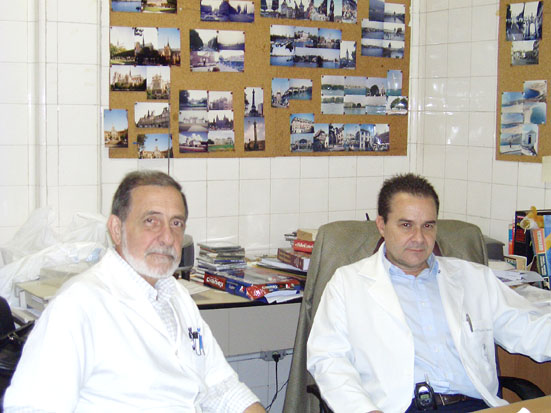
Dr. Waldemar Costa and Dr. Sampaio discussing research projects |
